This patient from Long Beach, Los Angeles sought help at the Dr. U Skin Clinic for eliminating a keloid cyst growth behind his right earlobe. The cluster of tissue had developed from a piercing and became quite large and noticeable. Due to treatment injections, an embedded cyst had developed within the keloid. Surgery was the only way to completely remove the bump. A surgical excision with flap closures was performed to prevent recurrence. In the front of the earlobe, a specialized surgical stitching technique was done to help render a more normal looking earlobe in the end.
What is a Keloid Cyst
A keloid cyst results from the overgrowth of keloid scar tissue which, in turn, has also developed a sac-like, fluid-filled cavity known as a cyst.
Keloids are a form of raised scar tissue made of thick, fibrous collagen. They grow excessively following an injury to the skin, such as an earlobe piercing. Other causes may include burns and surgical treatment.
The formation of collagen scars is a normal part of the skin repair process. But for those who are susceptible to developing keloids, the scar tissue keeps proliferating.
Keloids are benign growths. But in some people, they may be painful or itchy.
Patient’s Keloid Cyst in the Ear Before Dr.U’s Surgery
This patient has a history of keloid scarring. He underwent several unsuccessful attempts to treat his earlobe keloid with steroid injections. The purpose of such drugs is to help reduce the size of the tissue mass by breaking the bonds between the collagen fibers, reducing the amount of scar tissue below the skin.
For this particular case, the use of drug injections did not produce significant improvement. The patient then decided to have the growth surgically removed at the Dr. U Skin Clinic.
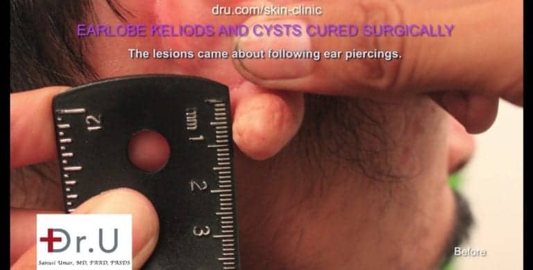
Here is a photo showing the keloid cyst growth behind the patient’s right earlobe. It measures about 1.5 cm in diameter.
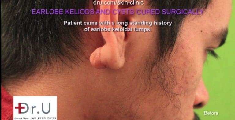
Due to its large size, the keloid tissue on the back of the patient’s earlobe had the tendency to hang downwards. He constantly had to reposition it to prevent it from showing.
With surgical intervention, he would no longer have to constantly worry about other people noticing the keloid.
Dr. U’s Keloid Cyst Removal Procedure
This excision surgery not only focused on the obvious goal of removing the keloid cyst growth but also two other important considerations:
- Preventing recurrence
- Restoring a normal looking appearance for the earlobe
Removing the keloid tissue cluster left a large open wound. However, simply stitching these edges together would incur a high risk of producing a new keloid growth as the skin tries to repair itself. The edges produce chemical signals which induce the formation of scar tissue. Patients who are already prone to developing keloids are then likely to struggle with new forms of unwanted collagen growth.
In some cases, individuals are instructed to wear medical earring pressure clips to prevent keloid recurrence. These devices must be worn for about twelve hours a day for 6-18 months.
Instead, we applied a flap wound closure method. This involved taking a surface section of the keloid tissue and stitching it in place to close the wound.
This prevents the signaling of scar formation which leads to recurrence. Also, using a flap instead of simply closing the wound helps preserve the original surface area. This prevents deformity of the earlobe in the final results.
For the front part of the earlobe, we used a specialized stitching technique which gathered the tissue together in the center, leaving a normal looking curvature at the base which is comparable with the other ear.
Without this approach, the bottom of the earlobe would have extended further downward, leaving an unusual looking appearance.
Before and After Photos of Earlobe Keloid Cyst Removal
Here are photos showing the patient’s earlobe before and after his keloid cyst excision performed at Dr. U Skin Clinic. Results are shown two months following his procedure.
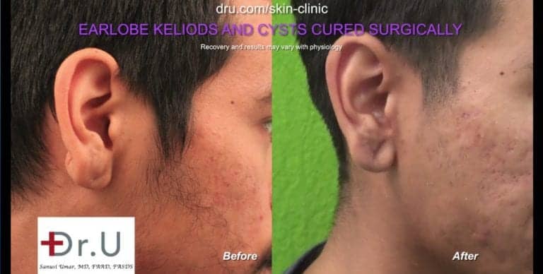
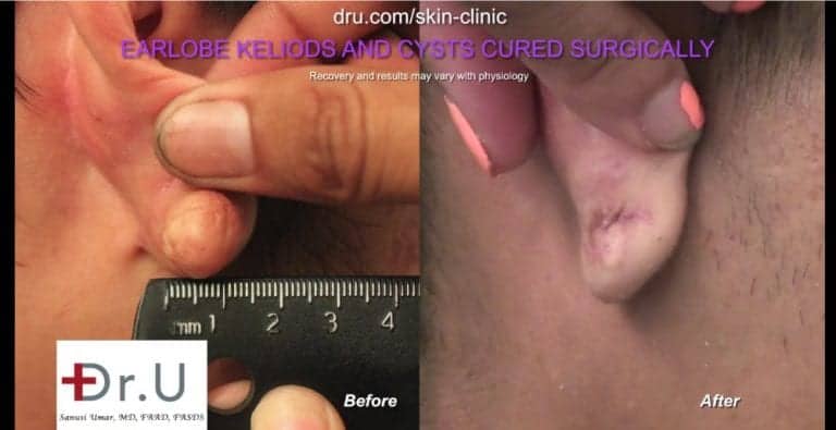
In these photos, it is clear that the right ear is now similar in appearance to the untreated left ear.
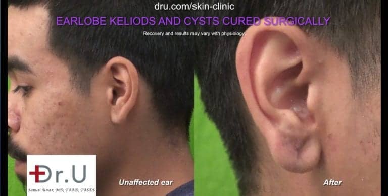
The patient’s wound healing results show no abnormal keloid scarring.
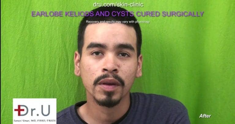
Keloid Cyst Excision in the Earlobe – Video of Long Beach, Los Angeles Patient
Watch this video to learn more about this patient’s keloid removal on his earlobe.
If you are interested in removing a keloid or cyst growth, use our complimentary online consultation form here:
Frequently Asked Questions – Keloid Cyst Removal
What causes a keloid or cyst to form on the earlobe?
There is usually a genetic predisposition behind the formation of keloid tissue or cysts. At this point, it is not clear why some people will produce excessive amounts of collagen in response to a skin injury. Scientists have yet to identify specific genes associated with the formation of keloid scars.
Earlobe cysts (also called epidermoid cysts or epidermal inclusion cysts) form when the rate of skin cell multiplication exceeds the rate of cellular shedding. This occurs below the surface in deeper layers of tissue and creates a sac or pocket filled with air, fluid or other substance.
Both keloids and cysts are benign forms of growth.
Is it really common for keloid scars to form as a result of ear piercings?
Due to the popularity of ear piercings and re-piercings, the formation of keloids has become a more prevalent cosmetic complication.
In one study, researchers documented that 42.3% of keloid cases in patients at a hospital were due to ear piercings.
At present, it is not clear if high incidences of keloid formation from ear piercings is due to the fact that the earlobes are a commonly pierced area, or if keloids from piercings are more likely to form than with other types of injuries.
What risk factors are associated with the formation of keloid scars on the earlobe?
Keloid scarring can occur in individuals of any race. However, people with darker skin tones face a much higher risk. Individuals of African, Hispanic, or Asian descent are 15 to 20 times more likely to develop keloids.
Also, keloids tend to develop in individuals younger than thirty years of age. Interestingly, though, a study conducted at the Medical College of Georgia found that children who received ear piercings below the age of eleven were less likely to develop keloids.
Pregnant women also face a higher risk of keloid formation as well as adolescent teens undergoing puberty.
How can a keloid in the ear be prevented?
A keloid in the ear can be prevented through an awareness of how prone you are to developing abnormal scarring. You may want to then reconsider having your ears pierced.
Another way to prevent keloid tissue from developing is by having the earlobes pierced much earlier in life, as the study by the Medical College of Georgia suggests.
One study found that earrings with metal backs may be a strong contributor to the formation of keloid on the back of the ear. Researchers have noticed that in keloid removal surgery involving ear piercings, most cases involve tumors present on the back of the earlobe, as opposed to the front.
The researchers concluded that metal earring backings may exacerbate a local “neurogenic” inflammation which contributes to the formation of large keloid scars.
Based on these findings, choosing earrings with non-metal backs may be an effective way to prevent keloids after getting the ears pierced.
Have more questions about keloid cysts on the ear or other locations? Use the button below to ask away.
Further Reading
Read about another earlobe keloid removal
Learn about Dr. U’s split earlobe repair procedures at the Dr.U Skin Clinic
