Acne Keloidalis Nuchae, a chronic inflammatory scalp condition, starts off as follicular pustule and papule bumps and later manifests as different lesion sizes, residing at various positions typically in the nape area, also known as the occipital area. Dr.U has outlined a breakthrough classification system for AKN lesions and proposes the use of specific selection criteria for matching patients with effective surgical removal and wound healing techniques. This article discusses his innovative methods for improving procedure outcomes for individuals struggling with large Acne Keloidalis Nuchae bumps.
The Need For AKN Patient Selection Criteria And Improved Surgical Methods
Although surgical excision is commonly performed to remove AKN lesions, current techniques remain limited in their ability to produce the most optimal cosmetic healing outcomes for patients. In many cases, the scarring outcomes and the shape of the final results are poor.
Dr.U recommends the use of objective screening criteria as necessary for selecting appropriate methods for specific wound sizes and positions on the occipital area.
Also, more innovative surgical methods are needed to improve the cosmesis of the final scar so that its appearance is as discrete as possible. Patients can then feel less self-conscious and confident enough to engage in normal, everyday interactions with others.
The Classification of Large Acne Keloidalis Nuchae Bumps
Some Acne Keloidalis Nuchae lesions have a vertical width greater than 3cm and located between the occipital protuberance and the posterior hairline. There are lesions within this category of large Acne Keloidalis Nuchae bumps which are large enough to breach both these borders. Others are less than or equal to 3cm. They may be located in the lower portion of the nape area, as opposed to the upper nuchal area.
In an article published by Plastic and Reconstructive Surgery (PRS) Global Open, entitled, Improved Outcome in Large Acne Keloidalis Nuchae Lesions With Innovative Surgical Approaches, Dr.U discusses novel surgical methods for removing both types of AKN lesions noted above. He proposes very specific techniques for each that would produce narrower scars that are linear in shape, aligned with a more natural looking posterior hairline. Previously, such outcomes were practically unobtainable, particularly for very large Acne Keloidalis Nuchae Bumps
Effective Surgical Methods Developed by Dr.U For Treating Large Acne Keloidalis Nuchae Lesions
Prior to the advent of Dr.U’s methods, a major limitation of existing AKN excisional techniques is that the final shape of the scar cannot be controlled for the most part, especially with larger, wider lesions. The most ideal outcome would be for the wound to close and result in an inconspicuous scar that is thin and straight. While this may be more feasible in cases where the AKN lesion is small and narrow, however, it becomes more difficult to achieve with bigger sized AKN formations.
The Bat Excision Technique Developed by Dr.U For Improved Posterior Hairline Scar
Dr.U has developed a method which he calls the Bat excision, intended to produce a straight linear scar aligned with the posterior hairline after the wound edges close. This involves excising the skin area surrounding the AKN lesion in the shape of a bat in a spread eagle position.
Combining Secondary Intention Healing With Bat Excision for Removing Large Acne Keloidalis Nuchae Bumps
Secondary intention healing is one of three main types of surgical wound healing. Instead of closing the edges immediately after excision is performed, the wound is left open, allowing the development of granulated tissue to fill in the empty space. A layer of new epithelium forms over this area to fill in the wound.
Within the context of Dr.U’s bat excision method, secondary intention healing also involves leaving the wound open. However, instead of allowing new skin tissue to form within the excised area, the top (superior) and bottom (inferior) edges will spontaneously contract to form a horizontal linear scar that defines a brand new posterior hairline.
Use of Tension Sutures for Large Acne Keloidalis Nuchae Bumps
An important component of Dr.U’s excisional method for larger AKN lesions (i.e. greater than 3cm) is the application of tension in specific directions. Bigger wounds will naturally have a greater distance between the top and bottom wound edges. Tension sutures help to facilitate the process of bringing them together.
Tension sutures generate contractile forces which promote collagen deposition and the formation of the extracellular matrix, rich in fibronectin. Furthermore, they also promote an optimal alignment of cell tissue which contributes to better aesthetics of the closed wound.
The application of tension sutures is also particularly useful in cases where the upper border of the lesion exceeds (i.e. superiorly) above the occipital notch. Tensional forces are then able to lower the wound below this point, encouraging better- looking results through secondary intention healing. The concave region beneath the occipital notch is a location that is more advantageous for healing due to the epithelization of granulation tissue.
As a final note, the degree of tension applied can be manipulated and adjusted along with various points of the sutures. Doing so helps to render the desired M-shape form that outlines the posterior hairline.
Debridement for Closing Wounds Resulting From the Removal of Large Acne Keloidalis Nuchae Bumps
During the wound closure process, contraction may slow or stop. This can be attributed to the formation of mature granulation tissue in the process of epithelization within the empty space of the wound. Dr.U’s recommends debridement, or the removal of this tissue to help the wound continue its course of contraction so that it is able to close to completion.
Using a 15-blade, curette or scalpel, the granulation tissue is curetted to a point right below the vertical wound edge. Neoepithelial tissue which forms in the margins is also excised. This helps to improve the definition of the wound margin. Using either electrocautery or aluminum chloride promotes the attainment of homeostasis.
According to Dr.U, debridement is an important step to take when granulation has occurred in wound margins that are still wide without much contraction progress due to premature epithelization processes. When combined with tension sutures, wounds can be prompted to close even when they seem to have stopped contracting for four weeks, or even more
Conditions That Promote Scar Stretching
Based on Dr.U’s empirical observations, scar stretching that exceeds 2,5cm is very likely to occur when the sagittal (i.e. horizontal) width of the lesion is 6.5cm or greater. A second contributing factor is constricted or reduced scalp laxity.
Dr.U’s Surgical Protocols for Tackling Two Types of Large AKN Lesions
Dr. U has outlined specific methodologies for treating two different types of large Acne Keloidalis Nuchae lesions, located in the lower or general nuchal region, at or near the posterior hairline.
Bat excision with SIH (Secondary Intention Healing) for AKN tissue growths less than or equal to 3cm in vertical width and located in the lower half of the nuchal area, between the occipital protuberance at the top border and the posterior hairline as the lower border.
Bat excision and Secondary Intention Healing With Tension Sutures For AKN lesions greater than 3cm in vertical width which also reside between the occipital protuberance and the posterior hairline. This approach also applies for lesions which are large enough to breach the two horizontal lines, but still within the general nuchal area.
These surgical methods, along with the use of debridement when needed allows the surgeon to exert control over the wound healing process along with a predictable timeframe. They also render a more natural looking posterior hairline in the form of a straight, horizontal line, an end result which, until now, has been challenging to achieve for lesions of bigger sizes.
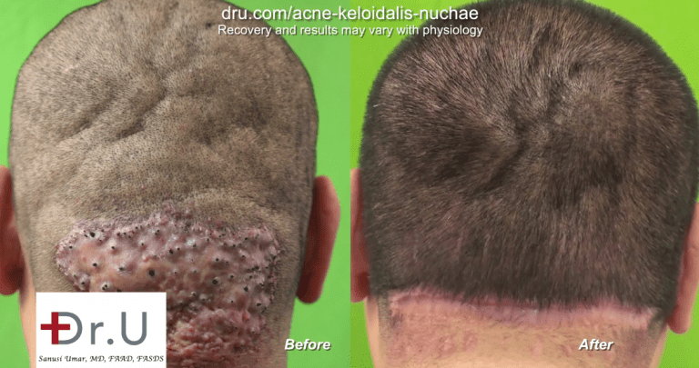
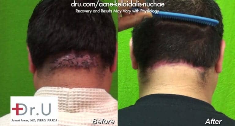
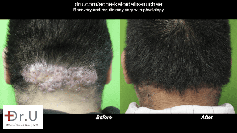
Research Study Outcomes for the Surgical Removal of Large Acne Keloidalis Nuchae Bumps
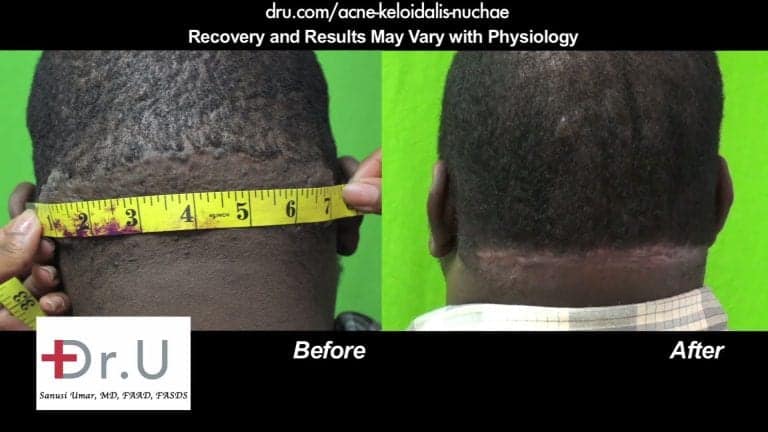
Dr.U observed the effects of his surgical methods on thirty-seven patients with Acne Keloidalis Nuchae, ranging in age from 25 to 58.2.
The bat excision was performed with an electrosurgical tip to create a bat-shaped wound.
- Three of these patients had smaller sized lesions, less than or equal to 3cm. Thus, their condition qualified for the application of the bat excision alone.
- In 35 of the 37 patients who underwent surgery for removing large Acne Keloidalis Nuchae bumps, the wounds fully contracted. Twelve of these individuals experienced small degrees of stretching where the final scar width remained less than 2.5cm.
- In two of the thirty-seven patients, the open wounds did not contract far enough to fuse the top and bottom wound edges. This was attributed to large sized sagittal widths (8 and 11cm) and very poor scalp laxity.
- Thirty-four patients had larger sized lesions which required bat excisions along with tension suturing. Four patients required debridement.
- In the four patients where debridement was necessary, three of these individuals required the use of tension suturing on a second round.
- In thirty-five patients, the wounds were able to spontaneously contact into either an M or U-shaped posterior hairline.
- Twelve of the thirty-five patients (32%) with fully contracted wounds experienced minor stretching of their scars, where the width was less than 2.5cm.
Dr.U observed that lesions greater than or equal to 6.5cm either resulted in scar stretching, greater than 2.5 cm or failure of the wound edges to merge and close.
In two of the patients, where wound closure had reached completion, hypertrophic scarring was observed in the final results. But this was easily and completely resolved through the use of steroid injections
Recurrence of AKN was not observed in any of the patients who underwent surgical treatment. When asked to provide their feedback about their procedures, 25 patients reported an average satisfaction score of 7.7 out ot 10, according to Likert scoring criteria at an average follow up time of 1.9 years following surgery.
If you are interested in speaking to Dr.U about the treatment and removal of Acne Keloidalis Nuchae lesions at any stage, click the free consultation button below and submit your information.
[dt_button link=”/acne-keloidalis-nuchae/free-consultation/” target_blank=”false” button_alignment=”center” animation=”fadeIn” size=”big” style=”default” bg_color_style=”default” bg_hover_color_style=”default” text_color_style=”default” text_hover_color_style=”default” icon=”fa fa-chevron-circle-right” icon_align=”left”]FREE CONSULTATION[/dt_button]Frequently Asked Questions – Large Acne Keloidalis Nuchae Bumps
What other surgical methods does Dr.U recommend for patients who wish to get rid of large Acne Keloidalis Nuchae bumps?
Patients with AKN wound lesions in the upper nuchal area which measure 3 cm or smaller in horizontal width qualify for the use of Trichophytic excision and closure techniques. This results in a very thin, discrete form of scarring where hair is encouraged to grow through and contribute to the concealment of the linear scar.
When are tension sutures usually removed in patients who undergo AKN surgeries with secondary intention healing in the context of bat excision?
Tension sutures are usually removed once they are no longer taut, at about two weeks
Why does debridement work to help wounds resume contraction in the surgical removal of large Acne Keloidalis Nuchae bumps?
It is likely that debridement helps to reset the cascade of wound healing processes that normally occur with a new wound.
Further Reading
