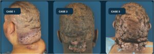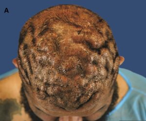

A newly published case series led by dermatologist and clinical investigator Dr. Sanusi Umar describes three men with the same combination of severe acne keloidalis nuchae, scalp-wide dissecting cellulitis, and extensive secondary cutis verticis gyrata. The findings support a candidate AKN-DCS-CVG syndrome that now requires independent validation.
By Sanusi Umar, MD
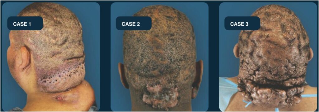
In a consecutive series of 12 men evaluated for suspected or confirmed dissecting cellulitis of the scalp, three met a demanding set of prespecified criteria. Each had clinicopathologically confirmed acne keloidalis nuchae at the posterior hairline and nape, dissecting cellulitis involving at least three scalp zones, and extensive cutis verticis gyrata involving at least three zones.
The repetition was not limited to appearance. It extended to anatomic distribution, patient-reported sequence, and biopsy findings. That convergence led our team to propose the AKN-DCS-CVG constellation as a candidate syndromic triad rather than three unrelated diagnoses occurring in the same patients.
The open-access paper, “A Candidate Syndromic Triad of Acne Keloidalis Nuchae, Dissecting Cellulitis, and Secondary Cutis Verticis Gyrata,” was published August 28, 2026, in Clinical, Cosmetic and Investigational Dermatology. The authors are Sanusi Umar, MD; Ochanya Ogah, MD; Jason Yang, MD; Belinda H Tan, MD, PhD; Nancy Aiead; and Paul K Shitabata, MD.
Three patients, one recurring clinicopathologic pattern
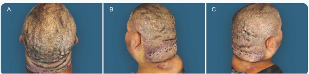
Acne keloidalis nuchae, or AKN, is a folliculocentric inflammatory scarring disorder that usually centers on the posterior hairline and nape. Dissecting cellulitis of the scalp, or DCS, produces boggy inflammatory nodules, drainage, and scarring alopecia. Cutis verticis gyrata, or CVG, describes deep ridges and furrows that give the scalp a folded appearance. In these cases, CVG was interpreted as secondary to severe chronic inflammatory and fibrotic scalp remodeling.
The three patients were 30 to 40 years old. Two identified as Hispanic and one as African American. All had class II or class III tumorous AKN, diffuse DCS, and extensive cerebriform scalp folding. Their nuchal and scalp lesions were not assigned solely from photographs. The diagnoses were supported by examination, trichoscopy, scalp-zone mapping, and biopsies reviewed by two board-certified dermatopathologists.
“These conditions can be diagnosed and treated separately, so their relationship may be missed,” Dr. Umar said. The practical concern is that an examination focused only on the most obvious lesion may fail to identify disease elsewhere on the scalp.
The nape disease was reported first
All three patients reported the same chronology. Disease at the posterior hairline and nape began first. Approximately 1 to 2 years later, diffuse inflammatory scalp disease and scalp furrowing were recognized.
That repeated sequence is important, but it must be interpreted carefully. The study was retrospective, and chronology depended on patient recall. It does not prove that AKN caused DCS or CVG. It does, however, identify a recurring order of clinical events that deserves prospective study.
The sequence also changes what clinicians should ask. A patient presenting with extensive scalp nodules and folds may need a detailed history of earlier papules, pustules, plaques, or masses at the posterior hairline. Conversely, a patient with severe AKN should be asked about new scalp pain, drainage, nodules, hair loss, and progressive folding outside the nuchal area.
CVG may mark a more extensive phenotype
The new report builds on an earlier 108-patient AKN cohort. In that study, CVG was present in three of seven patients with scalp-wide AKN, compared with one of 101 patients without scalp-wide AKN. In the three affected scalp-wide cases, AKN preceded CVG by approximately 1 to 2 years.
The current series adds a further element: all three patients with the complete triad had scalp-wide DCS supported by clinical and histopathologic assessment. This suggests that CVG may be a visible marker of a more extensive inflammatory and fibrotic scalp phenotype.
The finding does not establish a single mechanism. Secondary CVG may represent downstream remodeling produced by severe overlapping AKN and DCS rather than a separate disease process. That distinction is one reason the authors use the terms “candidate” and “syndromic triad” rather than claiming that a distinct inherited syndrome has been proven.
Multisite biopsy connected the clinical findings
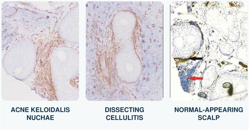
The biopsy framework sampled three diagnostic compartments in each triad case: a representative nuchal lesion, an active inflammatory scalp lesion, and clinically normal-appearing scalp located away from visible inflammation.
Nuchal specimens showed a fibrosing scarring alopecia centered on the folliculosebaceous unit, with follicular destruction, lamellar fibroplasia, mixed inflammation, keratinaceous debris, and foreign-body reaction, supporting AKN. Active scalp lesions showed follicular occlusion and rupture, neutrophil-rich suppurative inflammation, foreign-body reaction, and dermal fibrosis, supporting DCS.
The third compartment provided the most unifying observation. Clinically normal-appearing scalp in all three patients showed perifollicular infundibulo-isthmic lymphocytic inflammation and fibrosis, known as PIILIF. This pattern consists of lymphocytic inflammation and early fibrosis around the upper hair follicle, even where the scalp does not look visibly inflamed or scarred.
A companion 12-patient DCS study found PIILIF in all clinically normal-appearing scalp biopsies and in all sampled clinically inactive nodules. Active DCS lesions retained that pattern but added prominent suppurative inflammation and remodeling. Across AKN, DCS, and clinically normal-appearing scalp in the triad cases, immunohistochemistry also showed a similar CD4-predominant T-cell pattern and prominent CD117-positive mast cells.
These findings provide biologic coherence, but they are not disease-specific. The cross-sectional data cannot determine whether PIILIF precedes, follows, or coexists with clinically apparent AKN and DCS. It should therefore be viewed as a hypothesis-generating shared histologic signature, not a proven cause.
A father-son clue, but not proof of inheritance
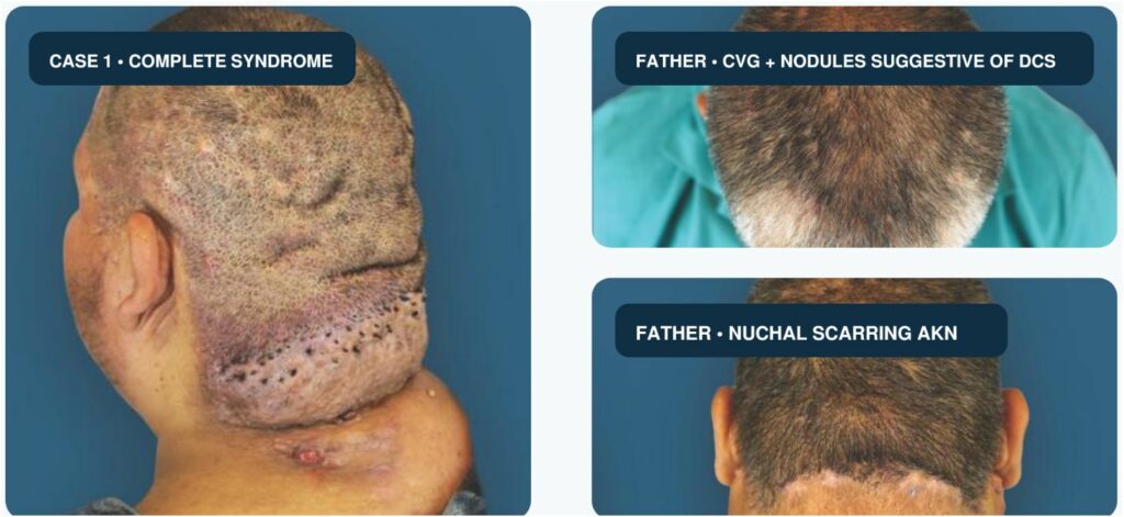
One patient’s father had severe cystic acne, chronic inflammatory boggy scalp lesions suggestive of DCS, diffuse scalp folding consistent with CVG, and a posterior hairline plaque suggestive of AKN. The visual similarity across two generations is notable.
It is not diagnostic proof. The father did not undergo biopsy, so the published report classifies his presentation as a clinically suspected but unconfirmed triad-like phenotype. The observation raises the possibility of shared susceptibility, especially because familial AKN and familial DCS have been reported, but one family cannot establish heritability or identify a genetic mechanism.
Systematic family histories and, where appropriate, clinical and pathologic evaluation of relatives should be considered in future studies. Any genetic conclusion will require a larger, independently ascertained cohort.
What clinicians should look for
The central clinical message is reciprocal examination.
In a patient with AKN, new scalp folds or diffuse boggy, painful, or draining nodules should prompt examination of the entire scalp for DCS. In a patient with DCS, the posterior hairline and nape should be deliberately examined for AKN papules, plaques, or masses. Extensive CVG in either setting may signal a high burden of inflammation and fibrotic remodeling.
Trichoscopy can help map perifollicular erythema, scale, casts, and other abnormalities that may not be obvious on routine inspection. When morphology is atypical, disease appears to overlap, or the diagnosis will affect management, trichoscopy-guided multisite biopsy can improve clinicopathologic adjudication.
The study did not evaluate treatment and does not establish that earlier therapy prevents DCS, CVG, scarring, or relapse. It therefore does not support a specific treatment algorithm. Its immediate value is diagnostic: recognize that disease may extend beyond the most visible region, examine the full scalp and nape, and consider early referral to a hair-disorders specialist.
A candidate syndrome that now needs validation
This was a small, retrospective, single-center case series drawn from a referral-enriched clinic population. The three cases identified among 12 men should not be interpreted as a prevalence estimate. Patient recall may have affected chronology, standardized longitudinal follow-up was unavailable, and the father’s triad-like phenotype was not histologically confirmed.
The next step is prospective, multicenter validation with standardized scalp-zone examination, trichoscopy, multisite biopsy, longitudinal follow-up, and systematic assessment of relatives. Larger studies can determine how often the constellation occurs, whether the reported chronology is reproducible, which patients are most at risk for extensive remodeling, and whether PIILIF has prognostic or therapeutic value.
For now, the evidence supports a careful conclusion: severe AKN, scalp-wide DCS, and extensive secondary CVG can occur as a reproducible clinicopathologic constellation. Recognizing that pattern may help clinicians identify coexisting disease across scalp regions sooner and may create a stronger foundation for future therapeutic research.
We are grateful to the patients who permitted publication of their clinical details and photographs, and to every coauthor whose work made the study possible.
View the Full Carousel on Instagram
View the Full Carousel on Instagram
Take the Next Step
Learn About Dissecting Cellulitis and PIILIF
Request an Acne Keloidalis Nuchae Consultation
Medical Disclaimer
| Medical disclaimer: This article is for educational purposes and summarizes an exploratory retrospective case series. It does not establish a diagnosis, prove causation, or provide individualized medical advice. Anyone with painful, draining, scarring, or folding scalp changes should seek evaluation from a qualified medical professional. |
Further Reading
- https://dru.com/acne-keloidalis-nuchae-dissecting-cellulitis-john-case/
- https://dru.com/piilif-in-dissecting-cellulitis-what-paired-biopsies-reveal-across-normal-appearing-scalp-quiet-nodules-and-active-lesionspiilif-in-dissecting-cellulitis/
- Umar, Sanusi et al. “A Retrospective Cohort Study and Clinical Classification System of Acne Keloidalis Nuchae.” The Journal of clinical and aesthetic dermatology vol. 14,4 (2021): E61-E67.
https://www.ncbi.nlm.nih.gov/pmc/articles/PMC8142833/